You are here
Orlando-Combita Heredia
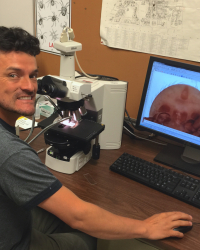
My research uses mites as model organisms to understand questions about behavior, evolution, and symbiotic relationships. Throughout my career, I've applied different tools such as molecular, morphological, and comparative methods to study evolutionary relationships among mites, in a systematics framework. I use a battery of microscopy techniques, including differential interference contrast microscopy (DIC), Confocal Laser Scan Microscopy (CLSM), Low-Temperature Scan Electron Microscopy (LT-SEM), Cryo Scanning Electron Microscopy (Cryo-SEM), Transmission Electron Microscopy (TEM), and Synchrotron X-ray microtomography (SR-mCT) to study sexual selection and feeding mechanisms in mites.
I am currently working with Dr. Alejandro Rico-Guevara as a Research Associate at the Behavioral Ecophysics, Department of Biology, University of Washington, gathering preliminary data such as specimen preparations, species identification and image acquisition for the project “Mites exploiting hummingbird-plant mutualisms: Behavioral and morphological mechanisms that enable a three-way interaction”. We are also working on the construction of 3D models through segmentation and reconstruction of internal organs and other structures processing based on CT scans of birds.
I am a Colombia Native. I earned a Biology Bachelor’s degree at the Universidad Nacional de Colombia in 2005. I studied insects and arachnids and my undergraduate research project on mites associated with Passalidae beetles under the direction of the national arachnid expert Eduardo Flores. I traveled to Venezuela to study for a few months with the mite specialist Magaly Quiroz at University del Zulia in 2004. I came to The Ohio State University (OSU) for the first time in 2006 and did the renowned Acarology Summer Program (soil mites). I came to OSU for the Acarology Summer Program in water mites in 2011. I did a Masters at the Universidad Nacional de Colombia on freshwater mites with the director of the invertebrate lab Dr. Rodulfo Ospina. My thesis project dealt with the diversity of aquatic mites and how this species could help answer questions about climate change and bioindication.
After earning a Master’s degree in 2013, I moved to the U.S. for a Ph.D. program at the Ecology, Evolution, and Organismal Biology department at OSU with Hans Klompen as my advisor. I studied mites associated with dung beetles and soil mites in tropical regions. During my Ph.D., I used a battery of light, electron, and X-ray microscopy techniques such as Confocal Laser Scan Microscopy (CLSM), Low-Temperature Electric Scan Microscope (LT-SEM), Cryo SEM, Fluorescence Stereomicroscope, Gyroscope, and Transmission Electron Microscopy (TEM). Additionally, I used different visualization and image edition programs such as FIJI, Cinema 4 D, Imaris, Drishti, MeshLab, 3D Slicer, and Adobe Suite tools, among others.
After being awarded my Ph.D. in 2020, I continued to work at the Acarology Lab with Hans Klompen. This position was part of a research grant in Uropodid mites and a collaboration that I established in 2020 between the Acarology Laboratory at Ohio State University (OSAL), the University of Washington (UW), and the Synchrotron facility at Karlsruhe Institute of Technology (KIT) in Germany. This grant aims to use internal morphology for systematic proposes. Through KIT, we gained access to Synchrotron X-ray microtomography (SR-mCT). Currently, with colleagues from the Universities of Washington and Colorado, we submitted a collaborative proposal to NSF. The current project aims to apply these techniques to study the ultrastructure of sensory receptors in mites associated with hummingbirds.

